Post by cebudentalimplants (@cebudentalimplants)
Language : EnglishTopics: Post-root canal painRoot canal infectionUnderstanding the Layers of Root Canal Complexity
Think of a tooth like a tree.
The crown is the top of the tree.
The roots are underground.
The canals are like tunnels running through the roots on your teeth.
Smaller accessory canals are like side branches just image vividly so it will be easy to understand.
A dentist must locate, clean, disinfect, and seal every significant canal. Missing even one infected canal can allow bacteria to survive and cause persistent infection.
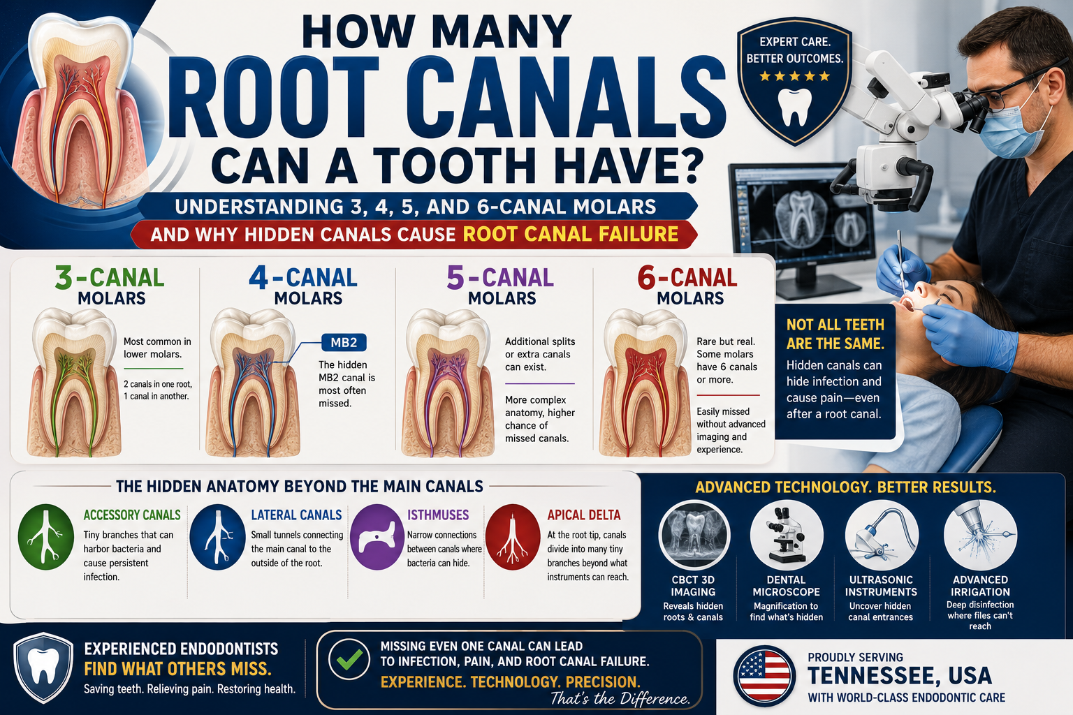
Level 1: Three-Canal Molars
Typical Anatomy
Most commonly found in:
Lower first molars
Some upper molars
Structure:
2 canals in one root
1 canal in another root
Total:
3 canals
Difficulty
Standard complexity
Usually predictable
Common Problem
Sometimes one canal is narrow or curved, making it difficult to clean completely. This make many season until it is finally complete. Skilled dentist knows and hand skills are years of experience of successfully done. This take time until finally complete
Level 2: Four-Canal Molars
Typical Anatomy
Very common in upper first molars.
Structure:
Mesiobuccal canal (MB1)
Hidden second mesiobuccal canal (MB2)
Distobuccal canal
Palatal canal
Total:
4 canals
Why Problems Occur
The MB2 canal is the most commonly missed canal in dentistry.
Many failed root canals involve:
A perfectly treated main canal system
One hidden MB2 canal left untreated
Bacteria remain inside that canal and continue causing infection.
Expert Insight
Modern endodontists using:
Dental microscopes
CBCT 3D scans
Ultrasonic instruments
find MB2 canals far more often than traditional methods.
Level 3: Five-Canal Molars
Typical Anatomy
Some molars develop:
Additional canal splits
Separate root systems
Complex anatomy
Total:
5 canals
Common examples:
Upper first molars
Lower molars with extra distal canals
Clinical Challenge
The canals may:
Curve sharply
Join together
Split apart multiple times
A dentist may locate four canals and still miss a fifth hidden pathway.
Level 4: Six-Canal Molars
Typical Anatomy
Rare but very real.
Examples include:
Lower first molars
Lower second molars
Upper molars with duplicated canal systems
Total:
6 canals
Why They Are Dangerous
A standard X-ray may show only four canals.
However:
Fifth canal hidden behind another root this needs advance technology so the fifth canal can be seen in x-ray so technology needed so it can bee seen?
Sixth canal buried deep within dentin
Without advanced imaging, these canals may remain undiscovered.
Beyond Canal Numbers: The Hidden Layers Most Patients Never Hear About
The number of canals is only part of the challenge.
Accessory Canals
Tiny side branches extending from the main canal.
They can contain:
Bacteria
Tissue debris
Infection
Some are too small to instrument directly.
Lateral Canals
Small tunnels connecting the main canal to the outside root surface.
Infection can travel through these pathways and create bone loss.
Isthmuses
An isthmus is a narrow connection between two canals.
Imagine:
Canal A
Canal B
Tiny bridge connecting them
Bacteria often hide in these bridges.
Many root canal failures occur because these areas are difficult to disinfect.
Apical Delta
Near the root tip, one canal may divide into many microscopic branches.
It resembles a river delta.
A dentist may clean the main canal successfully but bacteria can survive within these tiny terminal branches.
Why Experienced Endodontists Have Higher Success Rates
Modern specialists use:
1. CBCT 3D Imaging
Allows viewing:
Hidden roots
Additional canals
Curvatures
Bone infections
2. Dental Operating Microscopes
Magnification often exceeds:
8x
16x
25x
This helps identify canals invisible to the naked eye.
3. Ultrasonic Instruments
Used to uncover:
Calcified canals
Hidden canal entrances
MB2 canals
4. Advanced Irrigation Systems
Disinfect:
Isthmuses
Accessory canals
Apical deltas
Areas files cannot physically reach.
A Real-World Example
A patient in Tennessee arrived with pain six months after a root canal.
The original dentist believed the tooth had:
Four canals
A CBCT scan revealed:
A missed fifth canal
Infection around the root tip
Using microscope-guided retreatment, the endodontist:
Located the hidden canal.
Removed the old filling material.
Disinfected the entire canal system.
Sealed all five canals.
One year later:
Bone regenerated.
Infection resolved.
The tooth was saved.
Remember
A molar may contain:
3 canals (common)
4 canals (very common)
5 canals (complex)
6 canals (highly complex)
But the true challenge extends beyond the number of canals. Dentists must also manage:
Accessory canals
Lateral canals
Isthmuses
Apical deltas
Hidden canal branches
This is why an experienced USA endodontist often relies on microscopes, CBCT imaging, and advanced cleaning techniques. In many failed root canal cases, the issue is not the root canal itself—it is a tiny hidden canal or microscopic branch that was never discovered during the original treatment.
Can tooth extraction cause infection? Mild inflammation is normal, but increasing swelling, fever, or severe pain may indicate infection. Learn more about post-extraction infection warning signs in this detailed guide.
Book a consultation with our Cebu dental specialists for proper evaluation and care.
Ready to get expert guidance?
If you’re experiencing severe pain or delayed healing, book a consultation with our Cebu dental specialists to get proper evaluation and care.
Consultation or contact page .
Book a Consultation
For severe discomfort or delayed healing, book a consultation with our Cebu dental specialists today.
Looking for a dentist? Browse our Cebu dental directory to find trusted clinics near you.
Medical Review and Clinical Basis
This article is based on clinical dental guidelines and real patient recovery patterns observed after tooth extraction procedures. The information reflects common post-extraction healing stages, including normal clot formation, gum tissue repair, and signs of possible complications such as dry socket or infection.
While mild discomfort is expected after a dental extraction, worsening pain after Day 3, bad odor, exposed bone, or spreading pain may require professional evaluation. These symptoms are consistent with known post-extraction complications described in standard dental practice.
About Cebu Dental Implants
Cebu Dental Implants provides comprehensive tooth extraction, surgical procedures, and dental implant services in the Philippines. Our team evaluates post-extraction healing, manages complications such as dry socket, and advises patients on proper aftercare to prevent infection and delayed healing.
If you experience severe pain or unusual symptoms after extraction, early professional assessment is recommended to prevent further complications.
Important Medical Disclaimer
This content is for educational purposes only and does not replace professional dental diagnosis. Every patient heals differently. If symptoms worsen or do not improve within a few days, consult a licensed dentist for proper evaluation and treatment.
Author
This article was prepared by the Cebu Dental Implants content team in consultation with licensed dental professionals experienced in tooth extraction and implant procedures.
Source: https://cebudentalimplants.com/how-many-root-canals-can-a-tooth-have-hidden-canals-root-canal-failure
0 likes · 0 comments · 0 shares