Post by cebudentalimplants (@cebudentalimplants)
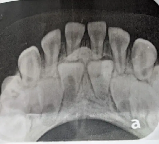
Image: Severity: CriticalTeeth Problems: Braces irritationGum infectionThe image appears to be an anterior dental occlusal radiograph, likely showing a developing/mixed-dentition situation. However, an X-ray alone cannot provide a definitive diagnosis or replace an in-person examination.
1. What I can see
The most important finding is crowding/overlap of the anterior teeth with developing or unerupted teeth positioned behind/among the erupted teeth.
Approximate tooth-by-tooth assessment
Area
X-ray appearance
Concern
Upper/lower anterior central region
Multiple overlapping tooth structures
Moderate–high concern for eruption/crowding problem
Central incisors
Roots/developing structures visible
Need assessment of eruption direction and root development
Lateral incisor regions
Crowding/overlap
Possible displacement/ectopic eruption
Canine regions
Developing tooth structures appear crowded
Need monitoring of eruption path
Bone around anterior teeth
No obvious large destructive radiolucency visible on this image
Reassuring, but this image is limited
Root areas
No obvious large apical lesion visible
Cannot exclude pathology without additional views
Gum/periodontal structures
Difficult to evaluate accurately from this projection
Clinical examination required
I do not see a large obvious abscess, destructive bone lesion, or massive cyst on this particular image. That is reassuring.
The bigger issue appears to be tooth position, eruption and crowding, rather than an obvious acute infection.
2. Important limitation
This is a single 2-D radiograph. There is considerable superimposition, so I would not diagnose a specific impacted tooth, supernumerary tooth, cyst, or orthodontic condition with certainty from this image alone.
For a definitive evaluation, a dentist/orthodontist may need:
Clinical examination
Panoramic X-ray
Periapical X-rays
Possibly a cephalometric X-ray
CBCT/3-D imaging only if clinically justified, particularly if an impacted tooth is close to another root or an important anatomical structure
A child's/adolescent's age is also very important because normal eruption timing changes the interpretation considerably.
3. If treatment is delayed for 14 days
This is where I would distinguish between orthodontic/eruption problems and infection.
If this is primarily an eruption/crowding problem
A 14-day delay usually does not suddenly cause major damage.
The problem is more likely to progress over months rather than days:
Crowding → abnormal eruption path → displacement/rotation → increasing difficulty creating space → potentially more complicated orthodontic treatment.
So 14 days is generally not an emergency purely because of the tooth positioning shown here.
If the patient develops infection
That is completely different.
If there is:
facial swelling
rapidly increasing gum swelling
fever
pus
severe spontaneous pain
difficulty swallowing
difficulty breathing
swelling spreading toward the eye/neck
then do not wait 14 days.
A dental infection can escalate much faster than an orthodontic problem.
4. Expected treatment/healing timeline
The exact treatment depends on what the dentist finds clinically.
Scenario A — retained baby tooth / creating eruption space
Examination and X-rays: same day
Extraction if necessary: usually one appointment
Gum healing: approximately 7–14 days
Underlying bone remodeling: several weeks to months
Permanent tooth eruption/position correction: months
Scenario B — orthodontic space creation
Diagnosis/planning: 1–3 appointments
Space creation: generally months
Tooth movement: commonly several months or longer
Retention afterward: required to stabilize the result
Scenario C — impacted tooth requiring surgical exposure
Surgical procedure: generally one appointment
Initial soft-tissue healing: roughly 1–2 weeks
Orthodontic traction/eruption: potentially several months
Final position: often significantly longer than 14 days
So if someone says "it will heal in 14 days," that generally means the soft tissue may heal substantially, not that the tooth-position problem itself has been completely corrected.
5. What could "scale up" if ignored?
For the radiographic appearance you provided, I would rank the potential progression approximately:
Current concern
→ anterior crowding/eruption abnormality
→ tooth displacement
→ worsening lack of space
→ rotation/ectopic eruption
→ possible root interference
→ more complicated orthodontic treatment
The highest concern is not the 14-day period itself. It is allowing an eruption/crowding problem to remain untreated for many months during active dental development.
If infection develops, however, the risk category changes immediately.
6. What I would do next
I would not extract anything based on this X-ray alone.
A reasonable clinical sequence is:
Step 1: Dentist/orthodontist examination
Step 2: Determine patient's age and eruption stage
Step 3: Obtain panoramic/periapical imaging if necessary
Step 4: Identify every permanent and primary tooth accurately
Step 5: Determine whether there is retained primary tooth, ectopic eruption, impaction, supernumerary tooth, or simply normal developmental overlap
Step 6: Assess available arch space
Step 7: Decide whether observation, extraction, space maintenance, or orthodontic treatment is appropriate
Step 8: Establish follow-up interval
My overall radiographic impression
Orthodontic/eruption concern: MODERATE → potentially HIGH depending on age and clinical findings.
Obvious acute infection on this image: LOW.
Immediate life-threatening finding visible on this image: NONE apparent.
But the absence of an obvious lesion on this one radiograph does not rule out disease elsewhere.
7. Finding a Cebu dentist through your directory
Your CebuDentalImplants.com Map Dental Clinic specifically provides a location-based directory for finding dental clinics.
Open the CebuDentalImplants.com Dental Clinic Map
The directory currently includes clinics in Cebu City, Mactan/Lapu-Lapu and Talisay, among other locations. For example, it lists D&G Dental Clinic, Ruflo Dental Clinic, Healthy Smile Dental Center–SM Seaside, ESL Dental Clinic Tabunok, and Almario Dental at Ayala Central Bloc.
Source: https://cebudentalimplants.com/dental-x-ray-case-analysis-severe-tooth-crowding-eruption-problems-and-what-happens-if-treatment
0 likes · 0 comments · 0 shares