Post by cebudentalimplants (@cebudentalimplants)
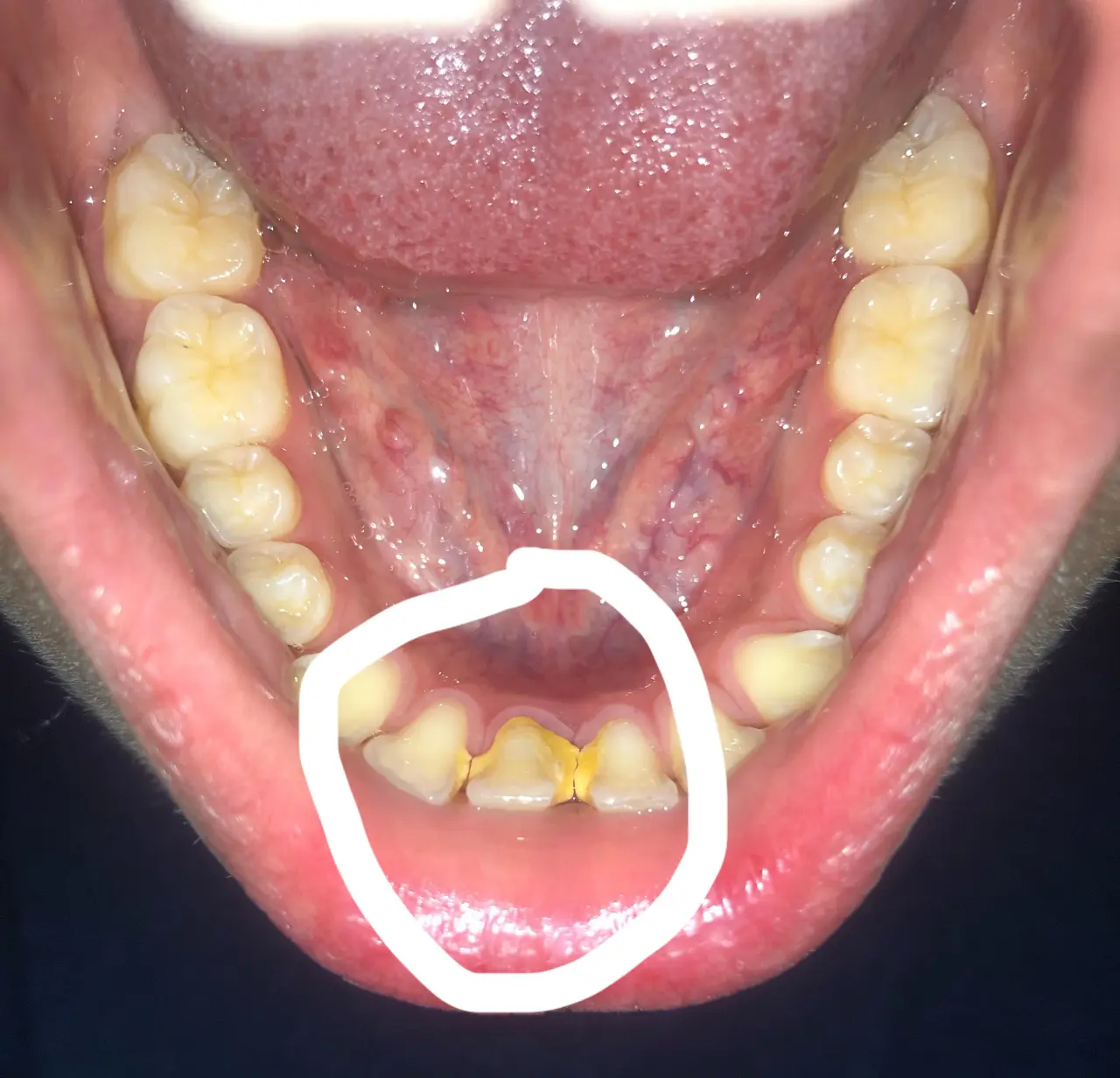
Image: Severity: ModerateTeeth Problems: Calculus casesI examined the photograph at 100% resolution and focused especially on the circled lower-front region. This is a clinical-photo assessment, not a definitive diagnosis; periodontal probing and appropriate X-rays are needed to determine whether there is bone or attachment loss.
Overall impression
The circled lower anterior teeth have a very heavy, hard-looking calculus/tartar deposit, especially around the gumline and between the lower incisors. The teeth are also noticeably crowded/rotated, which creates plaque-retentive areas.
The most important question is whether this is severe gingivitis only or whether it has progressed to periodontitis with periodontal pockets and bone loss. A photograph cannot establish that distinction. NIDCR notes that periodontal diagnosis requires gum measurements/probing, tooth examination and, when indicated, X-rays to evaluate bone loss.
Visual assessment
Finding
My assessment from the photo
Lower anterior calculus
Very heavy
Plaque retention
High
Lower incisor crowding/rotation
Moderate–severe
Gingival inflammation
Likely present
Gum recession
Possible; needs clinical measurement
Periodontal pockets
Cannot determine photographically
Bone loss
Cannot determine photographically
Abscess/pus
No obvious abscess visible
Obvious emergency
Not apparent from this photograph
The circled teeth
The central lower teeth appear to have large yellow/brown mineralized deposits extending along the cervical/interproximal surfaces. Some of what appears to be tooth structure may actually be calculus, so I would not assume that all of the visible yellow material represents damaged tooth.
The crowding is important because the overlapping surfaces are difficult to clean. Plaque that remains can mineralize into calculus, and calculus cannot be removed effectively by brushing or flossing; professional instruments are required.
Could this already be periodontitis?
Possibly, but I would not label it periodontitis from this photograph alone.
The dentist should measure periodontal pockets around every lower anterior tooth. Healthy pockets are generally around 1–3 mm; deeper pockets can indicate periodontal disease. X-rays can then determine whether supporting bone has been lost.
If there is attachment/bone loss, the treatment becomes more than a routine cleaning.
What I would do
Stage 1 — Examination
At the first appointment:
Full dental examination
Periodontal charting/probing
Check bleeding on probing
Check mobility of the lower incisors
Evaluate gum recession
Evaluate the amount and location of calculus
Take appropriate dental radiographs if periodontal disease is suspected
Determine whether this is gingivitis or established periodontitis
Stage 2 — Remove the calculus
If there is no significant attachment loss, professional scaling/debridement may be sufficient.
If periodontal pockets and attachment loss are found, the dentist may recommend scaling and root planing (SRP). ADA guidance describes SRP as a treatment for periodontal disease involving removal of plaque/calculus from tooth and root surfaces below the gumline.
Do not attempt to scrape this calculus off at home with metal instruments.
Stage 3 — Re-evaluation
After treatment, the dentist should reassess:
inflammation
bleeding
pocket depths
tooth mobility
gum position
remaining calculus
patient's home-care technique
If deeper pockets remain, additional periodontal treatment may be necessary.
What happens over 14 days?
If the primary problem is gingivitis caused by plaque/calculus, the gums can begin improving after professional removal of the irritants and improved oral hygiene.
A reasonable expectation is:
Day 0: examination + professional cleaning/debridement
Days 1–3: possible tenderness, sensitivity and some bleeding
Days 4–7: inflammation should begin decreasing
Days 7–14: gums may look considerably healthier
Around 2–4 weeks: periodontal reassessment may be appropriate depending on the initial findings
After deep cleaning, ADA notes that discomfort can last a day or two and sensitivity may last up to about a week; the gums can initially remain tender or bleed.
But 14 days does NOT mean the whole problem is healed.
If there is already periodontal bone/attachment loss, 14 days cannot regenerate the lost supporting structures.
The goal of the first 14 days is primarily to reduce the inflammatory burden and establish effective plaque control.
What could "scale up" if treatment is delayed?
This is the progression I would be concerned about:
Heavy calculus
↓
Persistent plaque accumulation
↓
Gingivitis / bleeding gums
↓
Periodontal pocket formation
↓
Attachment loss
↓
Alveolar bone loss
↓
Gum recession / tooth sensitivity
↓
Tooth mobility
↓
Possible tooth loss
NIDCR specifically notes that untreated gum disease can progress from inflamed gums to destruction of the supporting tissues and bone, potentially resulting in loose teeth or tooth loss.
However, waiting 14 days is very different from leaving this untreated for months or years. Periodontitis generally progresses over a much longer timeframe, although the appropriate treatment should not be unnecessarily postponed. NIDCR notes that periodontitis can take months or years to develop.
My concern level
Calculus: HIGH
Plaque-retention/crowding: HIGH
Visible gum inflammation: MODERATE–HIGH
Possible underlying periodontal disease: NEEDS TESTING
Immediate emergency from the photograph: NOT APPARENT
The biggest unknown is bone loss. That cannot be reliably assessed from this photograph.
Cebu dental clinic
For this case, I would specifically look for a dentist who can perform periodontal evaluation + professional scaling, rather than choosing a clinic solely because it advertises implants.
Your directory can be used here:
CebuDentalImplants.com Dental Clinic Map
Because you haven't provided your exact barangay/location, I can't honestly call one clinic the "nearest." If you're in the Mabolo/Cebu City area, current nearby options include:
Near Dental Clinic - Mabolo
Prestige Dental Clinic - Mabolo
Joon Dental Family Care
Affinity Dental Clinics Cebu
One Nadela Dental Group - Cebu
What to tell the dentist
You can simply say:
"I have heavy calculus on the lower anterior teeth. Please check my periodontal pocket depths, gum recession, tooth mobility and whether I have bone loss before deciding whether I need regular scaling or scaling and root planing."
That will focus the appointment on the real clinical question: gingivitis versus periodontitis.
If there is facial swelling, fever, pus, rapidly increasing pain/swelling, difficulty swallowing, or difficulty breathing, don't wait for a 14-day healing period—seek urgent dental/medical evaluation.
Mabolo•Use precise location
Source: https://cebudentalimplants.com/severe-tartar-lower-front-teeth-hidden-gum-disease-bone-loss-risk-treatment-process-14-day-healing
0 likes · 0 comments · 0 shares